BartterSyndrome.com - The Homepage of Dr. Lyn Sutherland
Bartter's Syndrome continued

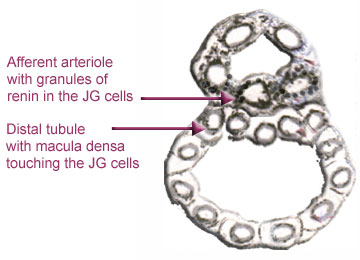
The last diagram shows how sodium, potassium and chloride are normally  transferred through the cells of the thick ascending limb and back into the blood.  In Bartter’s because the normal transfer is not going on, this abnormal load of electrolytes enters the distal tubule and the collecting ducts.  Even though the distal tubule itself is short it contains several types of cells and soon we discover the dire consequences of this overwhelming load.  We have entered a jungle and there be dragons ahead.

Distally the tubules are submerged by this surfeit of ions.  A large part of the sodium, potassium and chloride is lost to the body and the loss of potassium is made worse by the exchange of sodium for the body’s precious potassium under the influence of aldosterone.  Other changes occur as well but these will be discussed later. (We’re chicken.)

At this point it is imperative to go back to the JG cells and learn about renin and aldosterone and why Celebrex and similar drugs are so valuable.

In both Bartter’s, Gitelman’s and other electrolyte losing syndromes it is important to know the levels of renin and aldosterone in the blood.  Renin (pronounced Ree-nin) is synthesized and secreted by the juxtaglomerular cells of the kidney. (Rennin, pronounced Ren-nin is a product of a calf’s stomach and some of you may have eaten it as children, flavored and sweetened and called Junket.)

The study of Bartter’s and Gitelman’s was hindered until it became known that the cells of the thick ascending limb of the loop of Henle continued for a short way into the distal tubule.  It was now realized that the macula densa has the ascending limb’s cotransporters and channels just as described on the last page.  Therefore, if a patient has Bartter’s the cells of the macula densa no longer function normally.  Consequently, the juxtaglomerular cells are not receiving messages about the amount of sodium, potassium and chloride passing by.  The macula densa, no longer sensing salt, signals the JG cells to increase renin production and hence the lengthy process of salt retention begins.

Below is a diagram of the JG cells in the afferent arteriole touching the macula densa of the distal tubule.  (Look back to page 3 for orientation.)  The diagram has been kept very simple to avoid confusion with the efferent arteriole which has no JG cells and the glomerulus which is close by but is just as well out of the picture.

Jurgen Schnerman (2003) states that when sodium chloride is low at the macula densa, the macula densa cells begin to produce cyclooxygenase-2 or “COX-2” The COX-2 causes prostaglandin E2 or PGE2 to appear around the JG cells which promptly begin to synthesize and secrete renin.


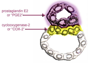
If we colour COX-2 yellow and PGE2 mauve the diagram above may be represented as below:


The PGE2 is not actually in the JG cells but in the interstitial tissue around them.  

Now you can see how Celebrex and similar anti-inflammatory drugs work. COX-2 is inhibited and so is the PGE2.  PGE2 cannot start the process of renin formation which leads to the production of aldosterone and many of the symptoms of Bartter’s syndrome vanish.  Sometimes these comparatively new drugs have side effects and in addition most or all patients must take a potassium supplement and often magnesium as well.

If you are wondering what happens when there is high salt at the macula densa, these cells become ATP/adenosine producing cells.

More about how the renin-angiotensin-system works next week.

[Previous Page]  [ 1 ]  [ 2 ]  [ 3 ]  [ 4 ]  [ 5 ]  [ 6 ]  [ 7 ]  [ 8 ]  [ 9 ]  [ 10 ]  [ 11 ]  [ 12 ]  [ 13 ]  [Next Page]
[ Lyn's Dedication Page ]